Dead on arrival
- By: Guy Hunt |
- Aug 11, 2008
- Share
- del.icio.us

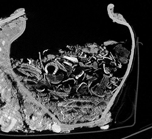
Medical scanner image through cremation urn 780. The urn is clearly visible (in cross section) as are three distinct layers of material within the urn.
We are very happy and excited to have the first scans back from two of our cremation urns which have been CT (computerised tomography) scanned using the latest medical technology at the City University. This is a brand new application of this technology in the world of archaeology.
The first cremation to be scanned was the cremation (Context: 780) featured in video episode 2. The whole cremation in its protective wrapping was put through the scanner in order to allow us to see inside before it is excavated in the lab. The results look amazing and we can make out different layers of bones within the remains inside the pot. Our human bone specialist Natasha is really happy with the results and she says that this will help us to excavate the cremation in a much better way than normal.
Unlike a modern cremations, Roman cremations don’t contain a ‘random sample’ of powdered bone, they contain the remains of a single individual. The body was typically burned on a pyre and afterwards the material is carefully collected and put into a container. Typically these are then buried, but they may also have been stored within above ground structures like a tomb or a Mausoleum.
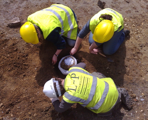
Liz, Nancy and Greg lifting the first cremation urn.
Overall I am very happy that we are showcasing yet another new technology at Prescot Street. The process itself is relatively cheap and hopefully it will save us valuable time in the lab while at the same time adding greatly to the archaeological information recovered. There will be more on this as the cremations are excavated.
I wonder if there is a record for the longest deceased patient that has ever been put through a medical scanner…
- (0)